
MIT researchers have developed a new method to convert skin cells directly into motor neurons without first turning them into pluripotent stem cells, a process that could make it easier to create neuron-based therapies for spinal cord injuries and neurodegenerative diseases such as ALS. Traditionally, cell conversion involves inducing skin cells to become induced pluripotent stem cells (iPSCs), which are then differentiated into specific cell types like neurons. While this method is widely used, it usually takes several weeks and doesn’t always result in mature, functional cells. The approach created by the MIT team gets rid of the iPSC stage entirely, offering a more direct and time-efficient route from skin cell to neuron.
The research, using mouse cells, focused on improving both the simplicity and efficiency of the conversion process. The team was able to produce more than ten neurons from a single skin cell. This level of yield is significantly higher than earlier methods of direct conversion, which typically achieved yields below one percent. According to Katie Galloway, the W. M. Keck Career Development Professor in Biomedical Engineering and Chemical Engineering at MIT and senior author of the study, this increase in efficiency allowed researchers to begin assessing whether the resulting neurons might be candidates for future cell replacement therapies.
Previous efforts by Galloway’s group involved delivering eight genes, six transcription factors and two proteins that stimulate cell proliferation, each with separate viral vectors. This made the process extremely difficult to control, especially when making sure that each gene was expressed at the right level in each cell.The study, published in Cell Systems, describes how the researchers were able to simplify the cell conversion process by narrowing down the number of necessary transcription factors. Instead of using six different factors, they found that just three, NGN2, ISL1, and LHX3, were enough to change skin cells into motor neurons when paired with two genes that encourage rapid cell growth. These two genes, p53DD and a modified form of HRAS, cause the skin cells to multiply quickly before starting the transition into neurons.
With only three transcription factors required, the team was able to deliver all of them using only one modified virus. This made it easier to have consistent gene expression across the cells. A second virus was also used to introduce the growth-promoting genes. Together, these two viruses worked in tandem to make the overall conversion process more efficient. The researchers noted that when skin cells were pushed into a state of rapid growth, they became more receptive to the reprogramming signals, which led to much higher neuron production.
The team also applied a similar method to human skin cells using an adjusted set of transcription factors. While the results weren’t as strong as with the mouse cells—the efficiency ranged from about ten to thirty percent—it still proved to be a promising direction. Importantly, this process was slightly quicker than the traditional method that involves generating and then differentiating pluripotent stem cells, taking about five weeks from start to finish.
In addition to improving the genetic approach, the researchers examined how different methods of gene delivery affected the efficiency of the conversion. In a second paper, also published in Cell Systems, they tested three types of viral vectors and found that retroviruses produced the highest conversion rates. They also discovered that reducing the density of the skin cells grown in culture dishes led to better outcomes. With these optimizations, the team achieved more than a one thousand percent yield of neurons from mouse skin cells in a process that took approximately two weeks.
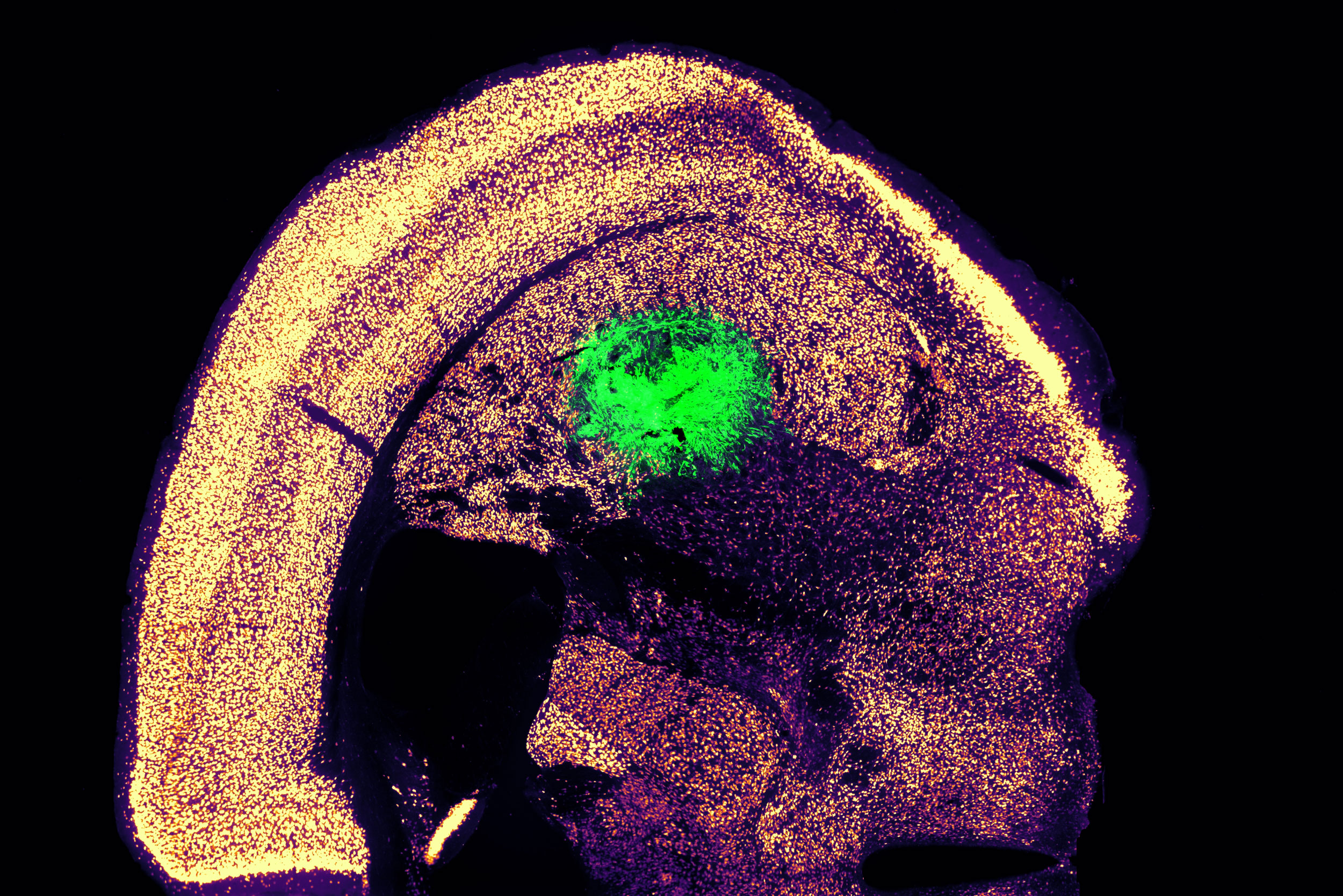
To find whether the newly created neurons could function in a living system, the researchers collaborated with colleagues at Boston University. They implanted the motor neurons into the brains of mice, specifically targeting the striatum, a region associated with motor control. Two weeks after implantation, the researchers found that many of the neurons had survived and had begun to integrate with the host brain tissue. In lab cultures, these neurons also showed signs of electrical activity and calcium signaling, suggesting they were capable of communicating with other neurons.


Leave a Reply